Internal Structure of Stems, Roots and Leaves
Table of Content |
|
|
Internal Structure of Dicot Stems
 Internal structure of a typical dicot stem shows following features:
1. Epidermis:
-
Epidermis is the outermost layer of the stem.
-
It is single layerd and lack of chloroplast.
-
Multicellular hairs (trichomes) and stomata are found on epidermis.
-
Outer side of epidermis a layer is present which is made up of cutin is called cuticle.
-
Epidermis plays a significant role in protection.
 2. Cortex:
In dicotyledon stem cortex divided into three parts:
 (a) Hypodermis: It is present just below the epidermis. It provides additional support to epidermis. It is thick multicellular layer. This layer is composed of collenchyma and their cells contain chloroplast. So hypodermis is green and photosynthetic.
(b) General Cortex: This part is composed 01 parenchyma. Storage of food is the main function of the cortex. Resin canal/ mucilage canal are present in it. These are schizogenous in origin. The innermost layer of the cortex is called endodermis.
(c) Endodermis: It is single celled thick layer. The cells of endodermis are barrel shaped. These cells accumulate more starch in stem of dicot. Thus it is known as ''Starch sheath".
3. Pericycle:
This layer is situated in between the endodermis and vascular bundles. The perieycle of stem is multilayered and made up of sclerenchyma. Sclerencymatous perieyle is also known as Hard bast.
4. Vascular Bundle:
The vascular bundles (wedge shaped) are arranged in a ring. Each vascular bund is conjoint, collateral and open. Each vascular bundle is made of phloem, cambium and xylem. Eustele is present in dicotyledon stems.
5. Pith:
This is well developed region, spreading from ring of vascular bundle to the centre. The cells of this region mainly made up of parenchyma.
Function of pith: Storage of water and food.
|
Cucurbita stem:
-
Angular stem is found in cucurbitaceae 'family.
-
It contains five ridges and five furrows. The vascular bundles arranged in two rows.
-
Each ring has five vascular bundles. In this way the total 10 vascular bundles are present.
-
The vascular bundles of outer ring are small in size and situated in front of ridges while the vascular bundles of inner rings are large in size and located below the furrows.
-
Vascular bundles are conjoint, bicollateral and open. Hypoderm is absent or less developed in furrows region and cortex contains chloroplast.
Internal Structure of Monocotyledon Stem
1. Epidermis: Epidermis is the outer most single celled thick layer. It is covered with thick cuticle. Multicellular hairs are absent & stomata are also less.
2. Hypodermis: Hypodermis of monocotyledor' stem is made up of sclerenchyma. It is 2-3 layered monocot stem rigidity is more in hypoderrrus where as in dieot stem elasticity is more. It provides mechanical support to plant.
3. Ground tissue: The entire mass of parenchyma cells next to hypodermis and extending to the centre is called ground tissue. There is no differentiation of ground tissue in monocotyledon stem. It means ground tissue is not differentiated into endodermis, cortex, Pericycle etc.
Note: Sometimes in some grasses, wheat etc. the central portion of ground tissue becomes hollow and is called Pith cavity.

4. Vascular Bundle:
-
Many vascular bundles are scattered in the ground tissue and V.B. are generally oval shape.
-
Vascular bundles lies towards the centre are large insize and-less in number.
-
Vascular bundles situated towards the periphery are small in size but more in number.
-
Each vascular bundle are conjoint, collateral and closed.
-
Vascular bundles surrounded by the layer of sclerenchymatous fibre are known as bundle sheath.
-
So vascular bundles are called fibro vascular bundles.
(a) Xylem: In xylem number of vessels is less. In metaxylem there occur two large vessels while in protoxylem there occur one or two small vessels. Vessels are arranged in V or Y shape. Just beneath protoxylem vessels, there occur a water cavity which is schizolysigenous in origin but major part of water cavity is lysigenous. This cavity is formed by disintigration of the element present below the proto xylem and neighbouring parenchyma.
Exception: In Asparagus water cavity & bundle sheath are absent.
(b) Phloem: It consists of sieve tube elements and companion cells. Phloem parenchyma is absent.
5. Pith: Pith is undifferentiated in monocotyledon stems. Atactostele is found in monocotyledon. This is highly developed stele.
Internal Structure Of Typical Dicotyledon Root

Internal structure of a typical dicotyledon root shows following features:
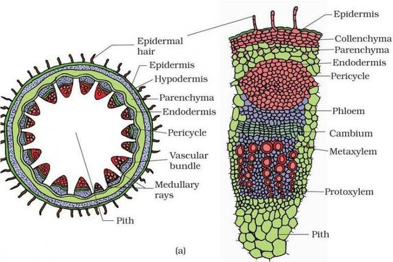
1. Epiblema: It is uniseriate outermost layer. It comprising tubular living components. Cuticle and stomata are absent. Unicellular root hairs are formed due to elongation of some cells of epiblerna. Epiblema also known as Rhizoderrnis or Piliferous layer. Root hair are present in maturation zone of root.
Note: Cells of epiblema which develop root hair called trichoblast.
2. Cortex: It is made up of parenchymatous cells.
Chloroplast is absent so they are nonphotosynthetic but chloroplast is present in roots of Tinospora and Trapa so they are photosynthetic.
Note: The cells of outer part of cortex -are suberized in old root. It is called exoderrnis.
Exoderrnis is found in some dicotyledonae roots and most of the monocotyledonae roots'.
3 Endodermis: This layer is situated between the pericycle and cortex. Casparian strips are present on radial and tangential wall of endodermis. These strips are made up of ligno suberin (mainly suberin).
Casparian strips are discovered by Caspari.
The cells of endodermis which are situated in front of protoxylem cells lack of casparian strips.
These are called passage cells/ transfusion cells. The number of passage cells is equivalent to the protoxylem cells. Passage cells provide path to absorbed water from cortex to pericycle.
|
4. Pericycle: It is single layered. It is composed prosenchyma.
Lateral roots are originated from the part of pericycle which is lying opposite to protoxylem. Thus lateral roots are endogenous in origin. A few mature cells of pericycle usually opposite to protoxylem, become meristernatic. These cells divide by periclinal divisions and form some layers of cells. These divisions are followed by anticlinal divisions forming a primordium which grows to form a lateral root.
|
5. Vascular Bundles: Vascular bundles are radial and exarch, xylem and phloems are separate and equal in number. The number of xylem bundles are two to six (diarch to hexarch).
But exceptionally, Ficus (Banyan tree) root is polyarch.
Parenchyma which is found between xylem and phloem is called conjunctive tissue.
6. Pith: In dicot root pith is less developed or absent.
Internal Structure of Monocotyledon Root

The internal structure of a typical monocotyledon root is similar to dicotyledon root:
(1) Number of xylem bundles are more than six (Polyarch) in monocotyledon root (exceptionally the number of xylem bundles are two to six in onion).
(2) Pith is well developed in monocotyledon root.
Internal Structure of Orchid Root
-
Velamen: These are found in aerial or hanging roots of some epiphytes (eg. orchid).
-
These are examples of multilayered epidermis.
-
These are present outside the exodermis
-
They absorb atmospheric moisture by imbibition.
-
Passage cells are found in both exodermis and endodermis in hanging roots of orchids.
Internal Structure of Dorsiventral Leaves

-
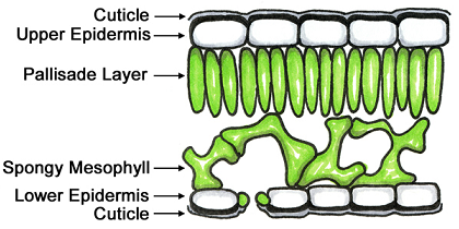
Cuticle is present on both surfaces but cuticle of upper surface is-more thick.
-
Dorsiventral leaves are mostly hypostomatic i.e. stomata present on lower surface.
-
In amphistomatic dorsiventral leaves stomata are more on lower surface as compared to upper surface.
-
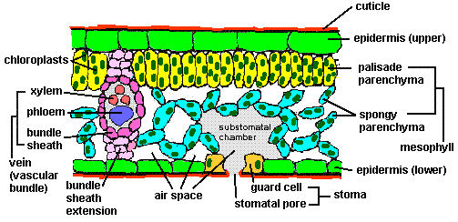
Mesophyll of these leaves is divided into two regions - Palisade tissue and spongy tissue.
-
Palisade tissue is found towards upper surface.
-
These cells have more chloroplasts and spongy tissue is found towards lower surface.
Internal Structure of Isobilateral Leaves
-
The thickness or cuticle on the both surface is equal.
-
Distribution of stomata on both surface are equal.
-
Isobilateral leaves are Amphistomatic i.e. stomata present on both sides.
-
Mesophyll of isobilateral leaves is not differentiated into palisade and spongy tissues.
-
It is completely made up of spongy tissues. Palisade tissue are absent.
Vascular Bundles of Leaves
-
Similar types of vascular bundles are found in both dorsiventral and isobilateral leaves.
-
Vascular bundles of leaves are conjoint, collateral and closed.
-
Protoxylem is situated towards the adaxial surface and protophloem towards the abaxial surface in the vascular bundle.
-
Leaves are devoid of endodermis and pericyde.
-
Vascular bundles are surrounded by a bundle sheath.
-
Bundle sheath in C-4 - plants is chlorenchymatous and remaining plants have parenchymatous or sclerenchyrnatous (mainly parenchymatous) bundle sheath.
Note:
1. Bulliform cells: Large cells are found in the epidermis of psammophytic (desert) grasses which are filled by liquid or empty (mostly) and colourless are called bulliform cells or motor cells. Upward folding of leaves during the sunny day takes place due to the presence of these specific cells. This is an adaptation to reduce the transpiration.
Example: Ammophila, Poa, Empectra and Agropyron etc. are Psammophytic grasses.
2. Epidermis of Nerium (both upper & lower) and Ficus (only upper epidermis) becomes multilayered. This is an adaptation to reduce transpiration.
Anomalous Primary Structure
(A) Anomalous structure in dicotyledon stem
 1. Scattered Vascular Bundles: In some of dicotyledon stem, vascular bundles are not arranged in a ring they are scattered in the cortex.
Example: Thalictrum, Numphaea. Papaver orientale & Peperomia.
II. Phloem on Innermost Radius: Generally phloem is situated in the ring of vascular bundles towards peripheral (outer) radius and xylem towards the inner radius. But anomalously in some plants the position of phloem is towards the inner side of xylem. Such type of phloem is called Internal or intraxylary phloem. Because, this. phloem lies towards the pith, so it is also known as medullary phloem.
This anomalous condition is found in Calotropis, Capsicum, Lepiadaenia. etc plants.
III. Medullary Vascular Bundles: In some plants vascular bundles are present in pith. These are found in addition to normal ring of vascular bundles. These are called medullary vascular bundles.
Example: Amaranihus, Boerhaaoia, Chenopodium, Mirabilis, Achyranthes, Bougainvillea, Raphanus sativus.
 IV. Cortical Vascular Bundles: Some of the vascular bundles are also present in the cortex of plants except the ordinary ring of vascular bundles. They are known as cortical vascular bundles.
Example: Casuarina, Nyctanthes and
V. Polystelic condition: Each vascular bundle is surrounded by a separate endodermis and pericycle in some plants. Hence each vascular bundle is a stele. It is the normal situation in pteridophytes but in some dicotyledons it is present abnormally.
Example: Primula, Dianihera
VI. Exclusively xylem vascular Bundle:
Abnormally, some vascular bundles are only formed by xylem except the normal vascular bundles.
VII. Exclusively phloem vascular Bundle: Abnormally, some vascular bundles are only formed by phloem except normal vascular bundles in some plants
Example-: Cuscuta & Ricinus communis
(B) Anomalous structure in monocot stem:
I. Vascular bundle situated in Ring: Normally vascular bundles are found in monocotyledon stem. It is in scattered form but in the stem of some monocotyledon plants vascular bundles are arranged in rings. Such as Triticum, Secale, Hordeum, Avena, Oryza etc. Members of family Gramineae
II. Eustele: Instead of atactostele, eustele is found abnormally in some monocotyledon stems. Presence of eustele is the basic feature of dicotyledons.
Example: Tamus communis.
|
Waiting meristem concept:
-
This concept was given by Buvat. According to this there is an inactive centre in the shoot apex which is known as waiting meristem and it acts as reservoir of active initials and on induction it give rise to reproductive apex.
-
Tannin is found ill latex of banana.
-
When it comes in contact with air it gets oxidised and becomes reddish brown in colour
-
Chewing gum is made by latex of Achrus sapota.
-
Tannin glands are found in camalia. These glands are. schizogenous in origin
-
Salt glands are found in Tamarix which secretes sodium chloride.
-
Chalk glands are found in plants of plumbaginaceal family which secretes calcium carbonate.
-
Multilayered (14 to 15 layers) epidermis is found in Peperomia.
-
Cystolith cells found in upper epidermis of Ficus leaf are called lithocytes. Cystolith are crystals of CaCO3·
-
The most durable wood is Tectona grandis.
-
Tracheids are the chief water transporting elements in gymnosperms.
-
Phloem is embedded into the secondary xylem in some plants. Such phloem is called included phloem or interxylary phloem. This is secondary anomalous structure.
Example: Leptadaenia, Salvadora etc. dicot stem.
Pericycle is absent in roots and stems of some aquatic plants.
In some monocotyledonae roots, pith is sclerenchymatous.
Example: Canna
In aquatic plants and ferns the epidermal cells and guard cells of stomata possess chloroplasts.


Q.1 - Monocot stem has
(a) Bicollateral closed vascular bundles (b) Bicollateral open vascular bundles
(c) Collateral open vascular bundles (d) Collateral closed vascular bundles
Q.2 - In monocot roots which types of vascular bundles are found
(a) Collateral, conjoint and closed (b) Radial V.B. with exarch xylem
(c) Bicollateral, conjoint and closed (d) Radial V.B. with endarch xylem
Q.3 - Abundant pith is characteristic of
(a) Monocot root and monocot stem (b) Monocot root and dicot stem
(c) Dicot stem and dicot root (d) Dicot root and monocot stem
Q.4 - Collenchymatous hypodermis is characteristics of
(a) Dicot stem (b) Monocot stem
(c) Monocot as well as dicot stem (d) Hydrophytes
Q.5 - Exarch and polyarch vascular bundles occur in
(a) Monocot stem (b) Monocot root (c) Dicot stem (d) Dicot root
Q.6 - The endodermis in dicot stem is also called
(a) Starch sheath (b) Mesophyll (c) Pili (d) Bundle sheath
Q.7 - Polyarch condition is seen in
(a) Monocot stem (b) Monocot root (c) Dicot root (d) Dicot stem
Q.8 - Which of the following is seen in a monocot root ?
(a) Large pith (b) Vascular cambium
(c) Endarch xylem (d) Medullary ray
Q.9 - Well developed pith is found in
(a) Monocot stem and dicot root (b) Monocot and dicot stems
(c) Dicot stem and dicot root (d) Dicot stem and monocot root
Q.10 - Sclerenchymatous sheath is present in vascular bundles
(a) Monocot root (b) Dicot root (c) Dicot stem (d) Monocot stem


|
Q.1 |
Q.2 |
Q.3 |
Q.4 |
Q.5 |
|
d |
b |
b |
a |
|
|
Q.6 |
Q.7 |
Q.8 |
Q.9 |
Q.10 |
|
a |
b |
b |
a |
d |
Related Resources:
-
Click here to refer the Useful Books of Biology for NEET (AIPMT)
-
Click here for study material on Structural Organisation in Animals
To read more, Buy study materials of Anatomy of Flowering Plants comprising study notes, revision notes, video lectures, previous year solved questions etc. Also browse for more study materials on Biology here.
View courses by askIITians


Design classes One-on-One in your own way with Top IITians/Medical Professionals
Click Here Know More

Complete Self Study Package designed by Industry Leading Experts
Click Here Know More

Live 1-1 coding classes to unleash the Creator in your Child
Click Here Know More

a Complete All-in-One Study package Fully Loaded inside a Tablet!
Click Here Know MoreAsk a Doubt
Get your questions answered by the expert for free
 In sunflower stem perieycle is made of alternate bands of parenchymatous and sclerenchymatous cells.
 Note:
Note: Note:
Note: Note:
Note: Note:
Note: Note:
Note: