Xylem and Phloem
Table of Content |

 Complex Tissues
A group of more than one type of cells having common origin and working together as a unit, is called complex permanent tissue.
The important complex tissues in vascular plants are :
(i) xylem and
(ii) phloem
Both these tissues are together called vascular tissue.
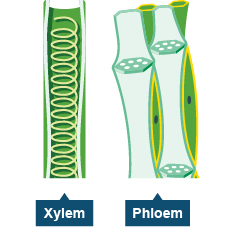
Xylem
-
The term Xylem is coined by Nageli.
-
The function of xylem is to conduct water & minerals salts upwards from the roots to stem & leaves and to give mechanical strength to the plan: body.
-
For conduction of water death of protoplasm if must. Dead tissues are more developed in water scarce conditions.
 On the basis of origin, xylem is divided into primary xylem and secondary xylem:
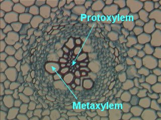
(i) Primary xylem originates from procambium.
(ii) Secondary xylem origtnates from vascular cambium.
(iii) Cells of protoxylem are small as compared to metaxylem
On the basis of development primary xylem is divided into two parts:
(1) Protoxylem
(2) Metaxylem
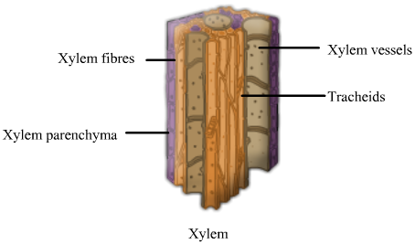
The Elements of Xylem
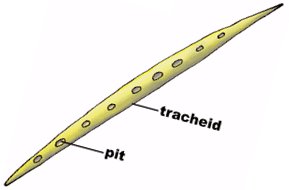
1. Tracheids
-
Tracheids are unicellular and having narrow lumen (but lumen of tracheids is wide than fibres)
-
Tracheids having a large lumen as compared to the fibres
-
Tracheids join together from their ends" to form a long rows.
-
These rows extending from the roots via stem to the leaves.
-
A transverse septum lies between each two tracheids. It bears pits. Water moves from one tracheid to another tracheid through pits.
-
Due to presence of transverse septum lumen is discontinuous in tracheids.
-
Tracheids are dead and lignified cells. The deposition of lignin on cell wall is responsible to form a different type of thickenings. (Pits are unlignified areas on lignified wall).
2. Vessels
-
It is advance conducting element -of xylem.
-
The basic structure of vessels is same as tracheids.
-
The lumen of vessels is wide than tracheids and end wall is perforated (Transverse septum is absent between two vessel elements. If present then porous). Thus vessels are more capable for conduction of water than tracheids.
-
Due to the presence of perforated end wall, vessels work as a pipe line during conduction of water.
-
Vessels contain usually simple pits at their lateral wall.
-
Thickening type-of wall is the same as tracheids.
3. Xylem Fibres
Xylem fibres provide strength to the tracheids and vessels. Mainly these fibres provide strength to the vessels. They are present more abundantly in secondary xylem.
4. Xylem Parenchyma
It’s cell wall is made up of cellulose. It store starch, fats and tannin etc.The radial conduction of water is the function of xylem parenchyma. (It conducts water to peripheral part of plant organ).Their walls possess pits.
Phloem
-
The term 'Phloem' is coined by Nageli.
-
The main function of the phloem is to conduct of food materials, usually from the leaf to other plant parts (eg. storage organ and growing regions)
-
On the basis of origin, phloem is classified into two categories primary and secondary phloem.
-
Primary phloem originates from procambium and secondary phloem originates from vascular cambiilrn.
-
On the basis of development primary phloem is categorised into protophloem and metaphloem.
-
The protophloem has narrow sieve tubes whereas metaphloem has bigger sieve tubes.
-
Phloem remains active for less duration as compared to xylem.
-
Phloem consists of 4 types of cells:
(i) Sieve cell → In Gymnospersm
(ii) Sieve tube → In Angiosperms
-
Sieve element was discovered by Hartig.
-
Sieve cells/sieve tube elements are living and thin walled.
-
A matured seive cell/sieve tube element lack nucleus. Thus these are enucleated living cells.
-
Central vacuole is present in each sieve cells/sieve tube element. Cytoplasm of sieve cells/sieve tube element show cyclosis in the form of thin layer around the vacuole.
-
In Angiosperm plants sieve tube elements are arranged with their ends and form sieve tube.
-
Sieve plate (oblique transverse perforated septa) is present between the two sieve tube elements at their end wall. It is porous. Materials are transported through these pores. Callose deposited on the radius of pores during dropping season (autumn) of leaves, to form a thick layer. This is called Callus pad.
-
Sieve plate is protected by callus pad. It also prevented from bacterial infection and drought.
-
Callose dissolves during spring season. Callose is b -1-3 glucan.
-
In Gymnosperms and pteridophytes, sieve cells do not form sieve tube and arranged irregularly. Sieve cell have sieve plates on their lateral walls. Thus conduction of food takes place in zig-zag manner.
-
In Angiosperms food conduction is erect and efficient.
-
Sieve elements contain special type of protein-P-protein (p-phloem).
|
1. Companion Cells
-
These are thin walled living cells. The sieve tube elements and companion cells are connected by pit fields present in their longitudinal walls, which is common wall for both and, with the death of one, other cell also dies.
-
A companion cell is laterally associated with each sieve tube element in Angiospermic plants.iln carrot more than one)
-
Sieve tube element and companion cell originates together. Both of them originate from a single mother cell. So called as sister cells.
-
The companion cells and sieve tube elements maintain close cytoplasmic connections with each other through plasmodesmata.
-
Companion cells is a living cell with large nucleus.
-
This nucleus also controls the cytoplasm of sieve tube element.
-
Companion cells are only found in Angiosperms.
-
Exception – Austrobaileya is angiosperm plant but companion cells are absent.
-
Special type of cells are attached with the sieve cells in gymnosperm and pteridophytes in place of companion cell. These cells are called as albuminous cells /strasburger cell.
2. Phloem Fibres
Fibres which are present in phloem are called Libriform fibres. These fibres are generally not found in primary phloem. These fibres provide mechanical support to the conducting elements (sieve cells and sieve 'tube),
3. Phloem Parenchyma
-
It is also known as bast parenchyma.
-
It's cells are living and thin walled. It store various materials. Eg. Resin, Latex, Mucilage etc.
-
The main function of phloem parenchyma is conduction of food in radial direction and storage of food. The conducting element of pholem is called Leptom, Leptom includes Sieve cells and Sieve tubes.
|
|
Special Tissue or Secretory Tissue
(I) Laticiferous Tissue
These are made up of long, highly branched and thin walled cells. These cells are filled with milky juice, called as Latex.
Latex is the mixture of saccharides, starch granules, alkaloids, minerals and waste materials.
Starch granules present in latex are dumble shaped.
Function:
(1) Latex provides protection to the plant.
(2) It prevents the plants from infection of bacteria and fungus. In latidferous tissue there are two types of cells-
(a) Latex vessels and,
(b) Latex cells
(a) Latex vessels:
-
These are articulated vessels. Latex vessels are formed due to dissolution of cells walls of meristematic cells. Thus these are syncytic cells as well as coenocytic (multinucleated).
-
Example: Latex vessels are present in Hevea, Ficus, Papaver, papaya, Argemone and Sonchus.
-
Highly developed latex vessels are found in the fruit wall of Poppy.
-
Opium is obtained from the latex, of Papaver somnijerum. It contains an alkaloid named as morphine.
-
An enzyme papain is obtained from the latex of papaya (Carica papaya).
-
Indian rubber is obtained from Ficus elastica and para rubber is obtained from Hevea bransiliensis.
-
Mostly latex is white in colour but in some plants latex is coloured.
-
In some plants latex is colourless Ex. Banana.
(b) Latex cells:
-
These are non articulated latex ducts/tubes. These are long, branched and multinucleated cells (coenocytic cells).
-
Example - Latex cells are found in Calotropis, Euphorbia and Nerium, Ficus religiosa
(ii) Glandular Tissue
-
As the name indicates this tissue is made up of glands.
-
These glands contain secretory or excretory materials.
Glandular tissues have two types of glands:
(i) Unicellular. Ex. - Urtica-dioica. These cells are present on the surface of the leaves. These are spiny glands in which formic acid is filled. It protects the plants from grazing animals.
(ii) Multicellular: Multicellular glands are of two types:
(1) External Glands: These are located on the surface of the plants and arising as an outgrowth from the epidermis. These glands are of various types:
(a) Glandular Hairs: They secrete gum-like sticky substances in tobacco and Plumbago, digestive juicy substance in Drosera.
(b) Nectar Glands: These glands secrete sugar solution. These are found in floral parts mainly in thalamus. These glands secrete nectar to attract the insects.
Exception: In Passiflora, nectar glands are found in leaves.
(2) Internal Glands: These glands are embedded in the tissues. Internal glands are of following types.
(a) Digestive glands: Digestive glands are found in insectivorous plants. These glands compensate the deficiency of N2 in insectivorous plants. These are found in Uiricularia, Drosera, Dionaea etc plants they secrete proteolytic juice.
(b) Mucous secreting glands: These glands secretes mucous. These are found in the leaves of betel.
(c) Oil glands: These secrete volatile oil. It acts as antiseptic. These glands are found in fruits & leaves of lemon & orange. Mostly, oil glands are lysigenous but in sunflower these glands are schizogenous.
(d) Water glands/Hydathode: These glands are related with guttation. Hydathodes are present in Tomato, Pistia and Eichhornia etc.
|


Q.1 - Vascular cambium and cork cambium are examples of
(a) Lateral meristem
(b) Apical meristem
(c) Elements of xylem and phloem
(d) Intercalary meristem
Q.2 - Laticiferous vessels are found in
(a) Xylem tissue
(b) Phloem tissue
(c) Cortex
(d) None of these
Q.3 - The cell wall of xylem cells is rich in
(a) Lipid
(b) Protein
(c) Lignin
(d) Starch
Q.4 - Tracheae, tracheids, wood fibres and parenchyma tissues are found in
(a) Xylem
(b) Phloem
(c) Cambium
(d) Cortex
Q.5 - Companion cells are usually seen associated with
(a) Fibres
(b) Vessels
(c) Tracheids


|
Q.1 |
Q.2 |
Q.3 |
Q.4 |
Q.5 |
|
a |
c |
c |
a |
d |
Related Resources
-
Click here to refer the Useful Books of Biology for NEET (AIPMT)
-
Click here for study material on Structural Organisation in Animals
To read more, Buy study materials of Anatomy of Flowering Plants comprising study notes, revision notes, video lectures, previous year solved questions etc. Also browse for more study materials on Biology here.
View courses by askIITians


Design classes One-on-One in your own way with Top IITians/Medical Professionals
Click Here Know More

Complete Self Study Package designed by Industry Leading Experts
Click Here Know More

Live 1-1 coding classes to unleash the Creator in your Child
Click Here Know More

a Complete All-in-One Study package Fully Loaded inside a Tablet!
Click Here Know MoreAsk a Doubt
Get your questions answered by the expert for free

 Note:
Note:  Note:
Note: 


 Note:
Note: Note:
Note:  Note:
Note: