Kingdom Monera
Table of Content |

 (1) Bacteria are the sole members of the Kingdom Monera.
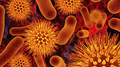
(2) Bacteria are grouped under four categories based on their shape: the spherical Coccus, the rodshaped Bacillus, the comma-shaped Vibrium and thespiral Spirillum.
(3) Compared to many other orgarusrns, bacteria as a group show the most extensive metabolic diversity.
(4) They may be photosynthetic autotrophic or chemosynthetic autotrophic. Some of the bacteria are autotrophic, i.e., they synthesise their own food from inorganic substrates.
(5) The vast majority of bacteria are heterotrophs, i.e.; they do not synthesise their own food but depend on other organisms or on dead organic matter for food.
Characteristics of Monera
Monera (Monos - single) includes prokaryotes and shows the following characters:
-
They are typically unicellular organisms (but one group is mycelial).
-
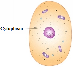
The genetic material is naked circular DNA, not enclosed by nuclear envelope.Ribosomes and simple chromatophores are the only subcellular organelles in the cytoplasm.
-
Mitochondria, plastids, golgi apparatus, lysosomes, endoplasmic reticulum, centrosome, etc., are lacking.
-
Sap vacuoles do not occur. Instead, gas vacuole may be present.
-
The predominant mode of nutrition is absorptive but some groups are photosynthetic (holophytic) and chemosynthetic.
-
The organisms are non-motile or move by beating of simple flagella or by gliding. Flagella, if present, are composed of many, intertwined chains of a protein flagellin. They are not enclosed by any membrane and grow at the tip.
-
Moneran cells are microscopic (1 to few microns' in length).
-
Most organisms bear a rigid cell wall (Peptidogl yean).
-
Reproduction is primarily asexual by binary fission' or budding.
-
Mitotic apparatus is not formed during cell division.
|
Bacteria Shape
-
Cocci: They are oval or spherical in shape. They are called micrococcus when occur singly as in Micrococcus, diplococcus
 when found in pairs as in Diplococcus pneumoniae, tetracoccus in fours, streptococcus when found in chains as in Streptococcus lactis staphylococcus when occurring in grape like clusters as in Staphylococcus aureus and Sarcine, when found in cubical packets of 8 or 64 , as in Sarcina.
 -
Bacilli: They are rod-shaped bacteria with or without flagella. They may occur singly (bacillus), in pairs (diplobacillus) or in chain (streptobacillus).
-
Vibrios: These are small and 'comma or kidney' like. They have a flagellum at one end and are motile, vibrio bacteria has curve in its cell e.g., Vibrio cholerae.
-
Spirillum: They are spiral or coiled like a corkscrew. The spirillar forms are usually rigid and bear two or more flagella at one or both the ends e.g., Spirillum, Spirochaetes etc.
-
Filament: The body of bacterium is filamentous like a fungal mycelia. The filaments are very small e.g., Beggiota, Thiothrix etc.
-
Stalked: The body of bacterium possesses a stalk e.g., Caulobacter.
-
Budded: The body of bacterium is swollen at places e.g., Rhodomicrobiu
Structure of Bacteria
 (1) Capsule: In a large number of bacteria, a slimy capsule is present outside the cell wall. It is composed of polysaccharides and the nitrogenous substances (amino acids) are also present in addition. This slime layer becomes thick, called, capsule. The bacteria, which form a capsule, are' called capsulated or virulent bacteria. The capsule 'is usually found in parasitic forms e.g., Bacillus, anihracis, Diplococcus pneumoniae, Mycobacterium tuberculosis.
(2) Cell wall: All bacterial cells .are covered by a strong, rigid cell wall. Therefore, they are classified under plants. Inner to the capsule cell wall is present. It 'is made up of polysaccharides, proteins and lipids.In the cell wall of bacteria there are two important sugar derivatives i.e., NAG and NAM (N-acetyl glucosamine and N-acetyl muramic acid) and besides L or D - alanine, D-glutamic acid and diaminopimelic acid are also found.
(3) Plasma membrane: Eachbacterial cell has plasma membrane situated just internal to the cell wall. It is a thin, elastic and differentially or selectively r permeable membrane. It is composed of large amounts of phospholipids, proteins and some amounts of polysaccharides but lacks sterols. It is characterised by possessing respiratory enzymes.
 (4) Cytoplasm: The cytoplasm is a complex aqueous fluid or semifluid ground substance (matrix) consisting of carbohydrates, soluble proteins, enzymes, co-enzymes, vitamins, lipids, mineral. salts and nucleic acids. The organic matter is in the colloidal state.The cytoplasm is granular due to presence of a large number of ribosomes. Ribosomes in bacteria are found' in the form of polyribosome.Membranous organelles such as mitochondria, endoplasmic reticulum, golgi bodies, lysosomes and vacuoles are absent. In some photosynthetic bacteria the plasma membrane gives rise to large vesicular thylakoids which are rich in bacteriochlorophylls and proteins.
(5) Nucleoid: It is also known as genophore, naked nucleus, incipient nucleus. There is nuclear material DNA which is double helical and circular. It is surrounded by some typical protein (polyamine) but not histone proteins. Histones (basic proteins) are altogether absent itt bacteria. This incipient nucleus or primitive nucleus is named as nucleoid or genophore.
(6) Plasmid: In addition to the normal DNA chromosomes many bacteria (e.g., E.coli) have extra chromosomal genetic elements or DNA. These elements are called plasmids. Plasmids are small circular double stranded DNA molecules. The plasmid DNA replicates independently maintaining independent identity and may carry some important genes. Plasmid terms was given by Lederberg (1952). Some plasmids are integrating into the bacterial DNA chromosome called episomes.
 (7) Flagella: These are fine, thread-like, protoplasmic appendages which extend through the cell wall and the slime layer of the flagellated bacterial cells. These help in bacteria to swim about in the liquid medium.Bacterial flagella are the most primitive of all motile organs. Each is composed of a Single thin fibril as against the 9+2 fibrillar structure of eukaryotic cells. The flagellum is composed entirely of flagellin protein.
(8) Pili or Fimbriae: Besides flagella, some tiny or small hair-like outgrowths are present on bacterial cell surface. These are-called pili and are made up of pilin protein. They measure about O.5-2mm in length and 3-5mm in diameter. These are of 8 types I, II, III, IV, V, VI, VII, and F types. I to F are called sex pili. These are present in all most all gram -ve bacteria and few gram +ve bacteria. Fimbriae take part in attachment like holding the bacteria to solid surfaces.The function of pili is not in motility but they help in the attachment of the bacterial cells. Some sex pili acts as conjugation canals through which DNA of one cell passes into the other cell.
Staining of Bacteria
 (1) Simple staining: The coloration of bacteria by applying a single solution of stain to a fixed smear is termed simple staining. The ells usually stain uniformly.
(2) Gram staining: This technique was introduced by Hans Christian Gram in 1884. It is a specific technique which is used to classify bacteria into two groups Gram +ve and Gram -ve. The bacteria are stained with weakly alkaline solution of crystal violet. The stained slide of bacteria is then treated with 0.5 percent iodine solution. This is followed by washing with water or acetone or 95% ethyl alcohol. The bacteria which retain the purple stain are called as Gram +ve. Those which become decoloutised are called as Gram -ve.
Differences Between Gram +ve Bacteria and Gram -ve Bacteria
S.No. |
Gram +ve Bacteria |
Gram -ve Bacteria |
|
1. |
They remain coloured blue or purple with gram stin even washing with absolute alcohol or acetone. |
The bacteria di not retain the stain when washed with absolute alcohol. |
|
2. |
The wall is single layered. Outer membrane is absent. |
The wall is two layered. Outer membrane is present. |
|
3. |
The thickness of the wall is 20-80nm. |
It is 8-12nm. |
|
4. |
The lipid content of the wall is quite low. |
The lipid content of the wall is 20-30%. |
|
5. |
The wall is straight. |
The wall is wavy and comes in contact with plasmalemma only at a few places. |
|
6. |
Merein or mucopeptude content is 70-80% |
It is 10-20%. |
|
7. |
Basal body of the flagellum has two rings of swellings. |
Four rings of swellings occur in the basal body. |
|
8. |
Mesosomes are more promiment. |
Mesosomes are less prominent. |
|
9. |
The bacteria are more susceptible to antibiotics. |
|
|
10. |
Fewer pathogenic bacteria belong to Gram+ve group. |
Most of the pathogenic bacteria are Gram* ve. |
|
11. |
Porins are absent. |
Porins or hydrophilic channels occur in outer membrane of cell wall. |
|
12. |
Cell wall contains teichoic acids. |
Teichoic acids are absent. |
Nutrition in Bacteria
On the basis of mode of nutrition, bacteria are grouped into two broad categories. First is autotrophic and second is heterotrophic bacteria.
-
Autotrophic bacteria: These bacteria are able to synthesize their own food from inorganic substances, as green plants do. Their carbon is derived from carbon dioxide. The hydrogen needed to reduce carbon to organic form comes from sources such as atmospheric H2, H2S or NH3.
-
Heterotrophic bacteria: Most of the bacteria cannot synthesize their own organic food. They are dependent on externalorganic materials and require atleast one organic compound as a source of carbon of their growth and energy. Such bacteria are called heterotrophic bacteria. Heterotrophic bacteria are of three typesParasites, Saprotrophs and Symbionts.
Arhaebacteria
 They are a group of most primitive prokaryotes which are believed to have evolved immediately after theevolution of the first life.
-
They have been placed in a separate subkingdom or domain of archae by a number of workers (e.g., woese, 1994).
-
Archaebacteria are characterised by absence of peptidoglucan in their wall. Instesd the wall contains protein and noncellulosic polysaccharised.
-
It has pseudomurein in some methanogens.
Mycoplasma (PPLO)
-
Mycoplasmas ar mollicutes are the simplest and the smallest of the free living prokaryotes.
-
They discovered in pleural fluid of cattle suffering from pleuropneumonia (nocard and roux, 1898).
-
The organisms are often called MLOs (pleuropneumonia like organisms).
-
The size ranges from 0.1-0.15um. a cell wall is absent. Plasma membrane forms the outer boundary of the cell.
-
Due to the absence of cell wall the organisms can change their shape and are pleomorphic.
Cyanobacteria
(Blue green algae, Cyanophyceae, Myxophyceae)
-
Cyanobacteria or blue – green algae are gram (=) photosynthetic prokaryotes which perform oxygenic photosynthesis.
-
Photosynthetic pigments include chlorophyll a, carotenoids and phycobilins.
-
Food is stored in the form of cyanophycean starch, lipid globules and protein granules.
-
Cyanobacteria evolved more than 3 billion years back.
-
They added oxygen to the atmosphere and paved the path for evolution of aerobic forms, including aerobic bacteria.
Difference Between Bacteria and Cyanobacteria
S.No. |
Bacteria |
Cyanbacteria |
|
1. |
The cells are comparatively smaller. |
The cells are comlarativey larger. |
|
2. |
The cell wll is 1-2 layered. |
The cell wall is four layered. |
|
3. |
Plasmodemata and pores do not occur in cell walls. |
They are often present. |
|
4. |
They exhibit lesser structural elaboration. |
They show higher degree of morphological complexity as well as structural elaboration. |
|
5. |
Bacteria are both autotrophic and heterotrophic. |
Cyanobacteria contain chlorophyll a as found in eucaryotic autotrophs. |
|
6. |
Autotrophic bacteria possess bacteriochlorophyll. |
Cyanobacteria contain chlorophyll a s found in eukaryotic autotrophs. |
|
7. |
Photosynthesis is anoxygenic. |
Photosynthetic is oxygenic. |
|
8. |
Photoautotrophic bacteria do not contain phycobilins. |
They possess accessory water soluble photosynthetic pigments known as phycobilins. |
|
9. |
Flagella may be present. |
Flagella are absent. |
|
10. |
Carbohydrate reserve food is glycogen. |
Carbohydrate reserve food is a special starch known as cyanophycean starch. |


Question1: Kingdom monera comprises the –
(1) Plants of economic importance
(2) All the plants studied in botany
(3) bldiryotic organisms
(4) Plants of Thallophyta group
Question2: According to Whittaker kingdom monera includes
(1) Unicellular eukaryotes
(2) Prokaryotes & akaryotes
(3) Slime molds & protozoa
(4) Multicellular & eukaryotes
Question3: Kingdom of unicellular eukaryotes –
(1) Monera
(2) Protista
(3) Fungi
(4) Plantae
Question4: Engler and Prantl created metachlarnydae to include –
(1) Polypetalous dicots
(2) Gamopetalous dicots
(3) Gamopetalous monocots
![]()

| Q.1 | Q.2 | Q.3 | Q.4 |
| 3 | 2 | 2 | 2 |
Related Resources
-
Click here to refer the Useful Books of Biology for NEET (AIPMT)
-
Click here for Study Material on Phylum- Chordata and Hemichordata
To read more, Buy study materials of Biological Classification comprising study notes, revision notes, video lectures, previous year solved questions etc. Also browse for more study materials on Biology here.
Watch this Video for more reference
View courses by askIITians


Design classes One-on-One in your own way with Top IITians/Medical Professionals
Click Here Know More

Complete Self Study Package designed by Industry Leading Experts
Click Here Know More

Live 1-1 coding classes to unleash the Creator in your Child
Click Here Know More


