Cell Envelope
Table of Content |
Cell Membrane
-
 The term was originally used by Nageli and Cramer (1855) for the membranous covering of the protoplast. The same was named plasmalemma by Plowe (1931).
 -
Plasmalemma or plasma membrane was discovered by Schwann (1838).
-
Membranes also occur inside the cytoplasm of eukaryotic cells as covering of several cell organelles like nucleus, mitochondria, plastids, lysosomes, golgi bodies, peroxisomes, etc.
-
They line endoplasmic reticulum, cover thylakoids in plastids or form cristae inside mitochondria. Vacuoles are separated from cytoplasm by a membrane called tonoplast.
Lamellar Models (= Sandwich Models)
They are the early molecular models of biomembranes. According to these models, biomembranes are believed to have stable layered structure.
The first lamellar model was proposed by James Danielli and Hugh Davson in 1935 on the basis of their physiological studies. Phospholipids form a double layer. The phospholipid bilayer is philic polar heads of the phospholipid molecules are directed towards the proteins. The two are held together by electrostatic forces. The hydrophobic nonpolar tails of the two lipid layers are directed towards the center where they are held together by hydrophobic bonds and van der Waals forces.
Mosaic Model
It is the most recent model of a biomembrane proposed by singer and Nicolson in 1972. According to this model, the membrane does not have a uniform disposition of lipids and proteins but is instead a mosaic of the two. Further, the membrane is not solid but is quasifluid. The quasifluid nature of the biomembranes is shown by their properties of quick repair, dynamic nature, ability to fuse, expand and contract, grow during cell growth and cell division, secretion, endocytosis and formation of intercellular junctions.

Membrane Transport
Passage of substances across biomembranes occurs by three methods- passive transport, active transport and bulk transport.
Passive transport
It is a mode of membrane transport where the cell does not spend any energy nor shows any special activity. The transport is according to concentration gradient. It is of two types’ passive diffusion and facilitated diffusion.
(1) Passive diffusion or transport across cell membrane. Here the cell membrane plays a passive role in the transport of substances across it. Passive diffusion can occur either through lipid matrix diffusion can occur either through lipid matrix of the membrane or with the help of channels.
 (2) Neutral solutes and lipid soluble substances. Neutral solutes and fat soluble substances can move across the plasma membrane through simple diffusion along their concentration gradient or from the side of higher concentration to the side of their lower concentration.
(3) Open channel transport. Membranes possess some open channels in the form of tunnel proteins. Water channels or aquaporins allow water and water soluble gases (CO2 and O2) to pass through according to their concentration gradient. Osmosis is an example of such a transport.
(4) Facilitated diffusion. it occurs through the agency of gated ion channels and permeases. Energy is not required. The transport is along concentration gradient.
(5) Ion channels are highly specific. There is a specific channel for each ion. Ions do not pass in dissolved state through ion channels but instead only ions move through them. Most ion channels are gated. Depending upon the stimulus required for opening the gated. More than 100 ion channels have been discovered. Movement through ion channels is according to concentration gradient. The rate of passage is quite high.
Active transport
-
 It is uphill movement of materials across the membrane here the solute particles move against their chemical concentration or electro-chemical gradient.
 -
Energy is required for the process. it is obtained from ATP.
-
Active transport occurs in case of both ions and nonelectrolytes, e.g., salt uptake by plant cells, glucose and phenolphthalein in case of renal tubules, sodium and potassium in case of nerve cells, etc.it is supported by various evidences
(i) absorption is reduced or stopped with the decrease in oxygen content of the surrounding environment.
(ii) metabolic inhibitirs like cyanides inhibit absorption.
(iii) active transport is also inhibited.by substances similar to solutes.
(iv) absorption of different substances is selective.
(v) cells often accumulate salts and other substances against their concentration gradient.
(vi) decrease in temperature decreases absorption. (vii) active transport is more rapid than diffusion. (viii) it shows saturation kinetics, that is, the rate of transport increases with increase in solute concentration not increase in solute concentration till a maximum is achieved.
Bulk Transport
-
It is transport of large quantities of micromolecules, macromolecules and food particles through the membrane.
-
It is accompanied by formation of transport or carrier vesicles. The latter are endocytotic and perform bulk transport inwardly. The phenomenon is called endocytosis.
-
Endocytosis is of two types, pinocytosis and phagocytosis.
-
Exocytic vesicles perform bulk transport outwardly. It is called exocytosis.
-
Exocytosis performs secretion, excretion and ephagy
(1) Pinocytosis: (Lewis, 1931).
-
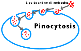
 It is bulk intake of fluid, ions and molecules through development of small endocytotic vesicles of 100 - 200 nm in diameter.
 -
ATP, Ca2+ fibrillar protein clathrin and contractile protein actin are required.
-
Fluid-phase pinocytosis is also called cell drinking.
-
After coming in contact with specific substance, the area of plasma membrane having adsorptive sites invigilates and forms vesicle. The vesicle separates. It is called pinosome.
-
Pinosome may burst in cytosol, come in contact with tonoplast and pass its contents into vacuole, form digestive vacuole with lysosome or deliver its contents to Golgi apparatus when it is called receptosome.
(2) Phagocytosis: (Metchnikoff, 1883).
-
It is cell eating or ingestion of large particles by living cells, .e.g., white blood corpuscles (neutrophils, monocytes), Kupffer's cells of liver, reticular cells of spleen, histiocytes of connective tissues, macrophages, Amoeba and some other protists, feeding cells of sponges and coelenterates.
-
As soon as the food particle comes in contact with the receptor site, the edges of the latter evaginate, form a vesicle which pinches off as phagosome.
-
One or more lysosomes fuse with a phagosome, form digestive vacuole or food vacuole. Digestion occurs inside the vacuole.
-
The digested substances diffuse out, while the residual vacuole passes out, comes in contact with plasma membrane for 'throwing out its contents through exocytosis or ephagy.
Cell Wall
-
 A non-living rigid structure called the cell wall forms an outer covering for the plasma membrane of fungi and plants.
 -
Cell wall not only gives shape to the cell and protects the cell from mechanical damage and infection, it also helps in cell-to-cell interaction and provides barrier to undesirable macromolecules.
-
Algae have cell wall, made of cellulose, galactans, mannans and minerals like calcium carbonate, while in other plants it consists of cellulose, hemicellulose, pectins and proteins.
-
The cell wall of a young plant cell, the primary wall is capable of growth, which gradually diminishes as the cell matures and the secondary wall is formed on the inner (towards membrane) side of the cell.
-
The middle lamella is a layer mainly of calcium pectate which holds or glues the different neighbouring cells together. The cell wall and middle lamellae may be traversed by plasmodesmata which connect the cytoplasm of neighbouring cells.
-
Pits 'are formed in lignified cell wall.
-
Tracheids in gymnosperms have maximum number of bordered pits.
-
In many secondary walls specially 'those of xylem the cell wall becomes hard and thick due to the deposition of lignin. With the increasing amount of lignin, deposition protoplasm is lost. First the lignin is deposited in middle lamella and primary wall and later on in secondary wall.
Growth of Cell Wall
By intussuception: As the cell wall stretches in one or more directions, new cell wall material secreted by protoplasm gets embedded within the original wall.
By apposition: In this method new cell wall material secreted by protoplasm is deposited by definite thin plates one after the other.
Cytoplasm
-
The substance occurs around the nucleus and inside the plasma membrane containing various organelles and inclusions is called cytoplasm.
-
The cytoplasm is a semisolid, jelly - like material.
-
It consists of an aqueous, structure less ground substance called cytoplasmic matrix or hyaloplasm or cytosol.
-
It forms about half of the cell's volume and about 90% of it is water.
-
It contains ions, biomolecules, such as sugar, amino acid, nucleotide, tRNA, enzyme, vitamins, etc.
-
The cytosol also contains storage products such as glycogenlstarch, fats and proteins in colloidal state.
-
It also forms crystallo - colloidal system.
-
Cytomatrix is differentiated into ectoplasm' or plasmagel (outer) and endoplasm or plasmasol (inner).
-
Cytomatrix is three dimensional structure appear like a network of fine threads and these threads are called microfilaments (now called actin filaments or microtrabecular lattice)' and it is believed to be a part of cytoskeleton. It also contains microtubules and intermediate cytoplasmic filaments.
-
Hyaloplasm contains metabolically inactive products or cell inclusions called deutoplast or metaplasts.
-
Cytoplasmic organelles are plastid, lysosome, sphaerosome, peroxisome, glyoxysomes, mitochondria, ribosome, centrosome, flagellum or cilia etc.
-
The movement of cytoplasm is termed as cyclosis (absent in plant cells).
Endomembrane System
The endomembrane system includes endoplasmic reticulum (ER), golgi complex, lysosomes and vacuoles. Since the functions of the mitochondria, chloroplast and peroxisomes are not coordinated with the above components; these are not considered as part of the endomembrane system.
|
Differences between SER and RER |
||
|
S.No. |
SER |
RER |
|
1) |
Ser does not bear ribosomes over the surface of its membranes. |
Red possesses ribosomes attached to its membranes. |
|
2) |
It is mainly formed of vesicles and tubules. |
It is mainly formed of cisternae and a few tubules. |
|
3) |
It is engaged in the synthesis of glycogen, lipids and steroids. |
The reticulum takes part in the synthesis of proteins and enzymes. |
|
4) |
SER gives rise to sphaerosomes. |
It helps in the formation of lysosomes through the agency of golgi apparatus. |
|
5) |
Pores are absent so that materials synthesized by SER do not pass into its channels. |
RER possesses narrow pores below its ribosomes for the passage of synthesized polypeptides into ER channels. |
|
6) |
SER is often peripheral. It may be connected with plasmalemma. |
It is often internal and connected with nuclear envelope. |
|
7) |
Ribophorins are absent. |
RER contains ribophorins for providing attachment to ribosomes. |
|
8) |
It may develop from RER. |
|
|
9) |
It has enzymes for detoxification. |
The same are absent. |
|
10) |
Vesicles for cis-face of Golgi apparatus are provided by SER. |
It provides biochemicals for Golgi apparatus. |
Related Resources
-
Click here to refer the Useful Books of Biology for NEET (AIPMT)
-
Click here for study material on Cell – the unit of life
To read more, Buy study materials of Cell: The Unit of Life comprising study notes, revision notes, video lectures, previous year solved questions etc. Also browse for more study materials on Biology here.
View courses by askIITians


Design classes One-on-One in your own way with Top IITians/Medical Professionals
Click Here Know More

Complete Self Study Package designed by Industry Leading Experts
Click Here Know More

Live 1-1 coding classes to unleash the Creator in your Child
Click Here Know More

a Complete All-in-One Study package Fully Loaded inside a Tablet!
Click Here Know MoreAsk a Doubt
Get your questions answered by the expert for free

