Internal structure of stem, roots & leaves: An important topic covered in AIPMT exam
Biology is not only about studying the external features of living organisms. Internal study is equally important. In medical entrance examination, you will surely get a question from internal study of different parts of plant and of animal as well. Here we are going to take a look to internal structure of stem, roots & leaves.
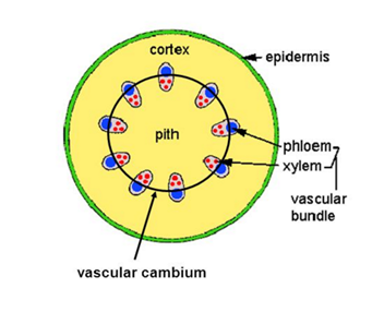
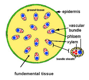
Stemis so responsible inside! Structurally monocot & dicot stems are quite different.
Let’s do a comparative study of both.
- Do not worry about the difficult terms. I have explained them at the end of article
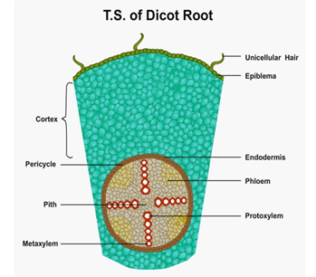
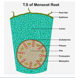
Do you know the root of roots?
I think it will be easy for you to learn the comparative study of roots as well.
So Guys! Want to study the comparative study of leaves as well?
Let’s do it by telling you the detail as well. Leaves are dorsiventral in dicots & isobilateral in monocots.
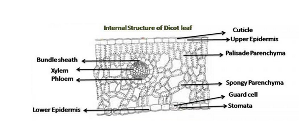
Dorsiventral leaves– To study these leaves, transverse section of mid rib in dicot leaf is studied.
The structures which are seen and studied in this leaf are:
- Upper epidermis – Single layered with thick cuticle on outside. Cuticle protects inner parts.
Epidermis controls transpiration rate. Cells are of parenchymatous type in it & they are without chlorophyll.
- Lower epidermis – Single layered with numerous stomata. Guard cells have chloroplast.
Stomata help in exchange of gases.
- Mesophyll – It is the ground tissue between upper& lower epidermis. Two types of parenchymatous cells are present.
- Palisade Parenchyma –Long & tubular cells. 2-3 layers are present. Photosynthesis occurs in it.
- Spongy Parenchyma – Cells are irregular in shape. Air cavities & intercellular spaces are present in it. They also show photosynthesis.
- Vascular Bundle – Larger towards the base of leaf blade. Its size is smaller towards leaf apex & margin.
Isobilateral leaves – Here also, transverse section of monocot leaf (wheat, maize etc.) is studied.
Structures observed are:
- Upper & lower epidermis – Both are single layered with stomata in it.
- Mesophyll – Only spongy parenchyma is present in it.  Palisade parenchyma is present only in few leaves.
- Vascular bundle – Sclerenchyma is present below & above the vascular bundle. Simple parenchyma is also present on the lateral side of vascular bundle.
I think; now doing a comparative study of both will be easy for you as I have provided all data here. Stillif you find any doubt or issue in it please post it on discussion board here. Your all queries will be answered J
And yes now you can take a look to some of the difficult terms used in my blog.
Conjoint: Here xylem and phloem lies in same bundle.
Collateral: Phloem is present towards the outer side and xylem lies toward the inner side.
Open: When Cambium is present between phloem and xylem.
Closed: Here Cambium is absent between phloem and xylem.
Exarch: When protoxylem lies towards the outerside&metaxylem is present towards the centre.
Endarch: When metaxylem occurs towards the outer side and protoxylem towards the inner side.
Prosenchyma– It is a plant tissue with elongated cells & tapering ends in it.
Thank you for showing interest in my blog. I am Anjali Ahuja, a member of askIITians family. We have created study material for AIPMT aspiring students. Click the link and buy whatever you like.

The article was really helpful. While preparing for the exams we at catalyser solved many years question papers and even the mock test had these questions. They taught us this well and the concept was made very clear to us.