The fine tuned design: Human Eye
The Fine Tuned Design: Human Eye
 Approximately one hundred billion (100,000,000,000) operations will have been already completed inside your eyes as soon as you finished reading the sentence with the speed of 36,000 bits of information every hour.
Do You Know?
- The information our eyes receive is sent to our brain along the optic nerve. This information is then processed by our brain and helps us make appropriate decisions
- Your eye is the second most complex organ after brain.
- You blink about 12 times every minute.
- 80% of our memories created through eyes.
- It’s impossible to sneeze with your eyes open.
- The cornea is the only tissue in the human body which doesn’t contain blood vessels.
- Your eyes contain around 107 million light sensitive cells.
 Advanced alarming system: Blink
Our eye is protected by the built-in early-alarming system. As the danger threatens, our nerves activate to engage the eyelids and stimulate the muscles that close the lids.
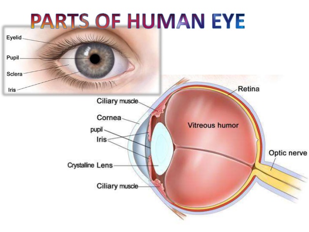
Blinking has two purposes – to keep the eyes lubricated and to protect the eye from foreign particles. It involves very complex processes and mechanisms with the help of several structures like retina, pupils, iris, lens, cornea, ciliary muscles and optic nerves. Proper assembly of these parts and cumulative effort helps the eye to function properly and makes our vision perfect.
Let’s have a glance over the structure of human eye
Human eye is the photoreceptor organ that receives light reflected by any object and sends the information to the brain through the optic nerve fibers.
Three layered-wall eye balls are fitted into the eye socket externally protected by eyelid.
Like the window in our home is meant for looking the outside world, the same way the cornea function is to focus incoming light, thus allowing it to pass through the lens towards the retina at the rear of the eye.
You must be wondering, if cornea do not contain blood vessels then from where its tissues get nutrients and oxygen?
Don’t panic!
The cornea gets nutrients from the tears and oxygen directly from the air.
Retina: The Photographic Film
Retina is the inner most layer of the eyeball. In human-eye, retina is inverted (sensory cells lie opposite to the entry of the light). The sensory cells, cone shaped and rod shaped, responsible for color vision in bright light and for black & white vision in dim light respectively.
Lens: Biconvex and Circular
The lens is made up of fibrous materials. Its position is fixed but the focal length is adjustable, this property of lens is called Power of Accommodation. The focal length of human eye lens is 1.5 cm, with refractive power of 66.7 Diopter at rest.
Mechanism of Vision:
So guys, ever thought about the process by which we see something?
Let me tell you about the mystery behind the fact.
Light rays enter our eyes through a series of stations viz., Conjunctiva, Cornea, Aqueous humour, Lens and Viterous humour and finally over Retina.
Retina acts as photographic film that of old versions cameras in which roll films were used. Retina contains sensory cells named cone shaped and rod shaped on the basis of their shape. These sensory cells absorbs light and activates the pigments present in the membranes of the vesicles they contain. The light then converted into action potentials in the membranes.
The action potentials travel as nervous impulse through the sensory cells and reach the synaptic knobs of the bipolar nerve cells. The bipolar nerve cells transmit the information to the ganglion and finally to the brain through optic nerves.
One interesting fact is that our retina receives inverted image of any object. It is our brain that analyzes and makes us see the erect images.
So, guys your feedback is crucial for our qualities to develop. You can comment below and you find the information worth informative and if you are curious to know more about the human eyes and how it is controlled by our nervous coordination, you can click this link.
Well ! I am Aabid Hussain, a member from of askIITians. We always try to give best of study material to our students. You can take a glance through this link if you want to check it out.


